Setting up the microscope

Bacteria are so small, they are on the limits of what we are able to see using a light microscope (why?). To get a clear image, we need to use the highest quality microscope and it MUST be correctly set up so that it is functioning at optimum efficiency:

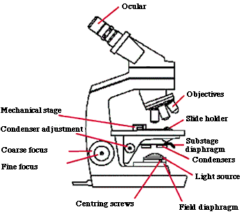
  1. With the substage condenser turret in the 0 position, using a X10 objective, focus on a mark on a microscope slide (make sure both iris diaphragms are fully open and that the light intensity is set mid-way).
  2. Stop down the field iris diaphragm and observe the light contract to a small circle in the field of view. If necessary focus the edge of the circle until sharp, using the condenser rack and pinion.
  3. If the circle of light is not central, adjust until central with centring screws beside the field diaphragm.
  4. Fully open the field iris diaphragm - the light setting is now correct for all objectives.
  5. All bright field objectives should be used with the substage condenser in the 0 position.

To set up the microscope for phase microscopy:

  1. Swing substage condenser to 40 and swing in X40 phase objective. Make sure that objective is nearly touching coverslip.
  2. Remove one of the eye pieces and insert phase telescope.
  3. Look down phase telescope and focus phase rings using the adjustment collars on the phase telescope. Make the two phase rings exactly concentric by moving substage condenser in light path. (The process has to be repeated for any other phase objectives and matching phase condensers).
  4. Remember that X40 phase has to be used with the substage condenser in the 40 position. It is a good idea to check the phase alignment each time you use phase contrast. When the rings are concentric, the microscope is in phase.
  5. Remove the phase telescope and replace the normal eyepiece before viewing slides.
see this on video

For the rest of this experiment, we will be using bright field microscopy.

Now let's look at some bacteria


DISCLAIMER          © AJC 2002.